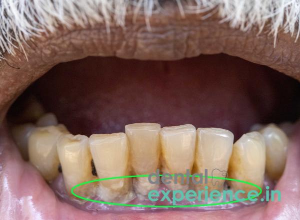
Age-related darkened teeth are defined as the natural and unavoidable changes that occur in tooth color and shade over years from the combination of intrinsic physiological changes occurring within the tooth itself and extrinsic stains accumulating on the surface of the teeth. The process of aging causes the outside transparent enamel to wear away through daily use of teeth for eating, drinking acids, and grinding action from toothbrushing. At the same time, secondary dentin forms beneath the enamel, becomes thicker, and takes on a darker shade of yellow or brown as minerals deposit into it. Since dentin is naturally darker than enamel, an increase in the amount of dentin due to an increase in thickness—combined with the decrease in the outer enamel—causes a shift in how light travels through the tooth, making the tooth look duller, yellower, grayer, or darker brown. Additionally, as secondary dentin is deposited throughout life, the center of the tooth becomes smaller since the space occupied by the original pulp has been reduced, causing less light to pass through the tooth. When dental classification systems list changes that occur to teeth over time due to aging, they fall into two categories: internal tooth discoloration (also called intrinsic dyschromia), which refers to darkening inside of the tooth, and extrinsic staining, which occurs from the accumulation of stain on the outside surface of the tooth.
Clinical Definition: Darkening of teeth due to loss of outer enamel and darkening of inner secondary dentin.
Major Visible Changes: Gradual darkening of many teeth at once due to uniform yellow, amber, brown, or grayish colors.
Possible Urgent Signs: Darkening of one tooth alone, sudden onset of sharp pain in a single tooth, temperature sensitivity in a single tooth, or inflammation around gums of a single tooth.
Most Common Restorative Procedures: Laser/LED bleaching done in office, microabrasion, composite resurfacing, porcelain laminates, full-coverage ceramic crowns.
Local Context: Extremely common in adults residing in Kerala, India, who have consumed large amounts of chromogenic hot drinks throughout their lives as well as chewed betel products and drank water high in mineral content.
Color Change: Teeth collectively undergo progressive color shifts from bright white or light off-white to uniform yellow, amber, dark brown, or gray shades.
Transparency Loss: The incisal edges (the biting edge) become dull and opaque rather than being transparent like glass.
Texture Changes: Fine cracks form on the enamel's surface, called craze lines, which can soak up darker pigments.
Clinical Crown Shortening: Attrition from years of chewing causes visible flattening of the biting surfaces of all teeth, making them appear shorter.
Exposure of Roots: Mild gingival recession exposes a part of the cementum covering the roots of the teeth.
Normal discoloration among most teeth as we grow older may be expected, but there are warning signs that warrant immediate dental care:
Discoloration Differences: When one tooth becomes very dark with a gray or black tint, this indicates internal pulp necrosis (death of the pulp nerve) from years ago due to a traumatic injury that was silent at the time.
Pain Due to Dentin or Root Caries: Aching pain within a tooth after it has turned dark, which could indicate internal inflammation or micro-leakage into the tooth's pulp chamber.
Sensitivity to Heat / Cold: Prolonged sharp pains when consuming either very hot coffee/tea or cold food/drink; sensitivity to heat/cold usually means dentin or root caries is exposed
Dentin Hyper-Thickening: Dentin is continually produced by the dental pulp to protect it throughout life. This secondary dentin production compresses the pulp chamber and increases the degree of inner opacity.
Enamel Micro-Thinning: Years of chewing stresses, abrasives used in toothpaste, and acid exposure all contribute to thinning of the enamel layer, ultimately exposing the dark dentin below.
Chromogen Staining: Daily dietary habits contain pigments that penetrate deep into the micro-cracks within the porous structure of the enamel, creating irreversible staining on the enamel. Frequent ingestion of dark tea, filter coffee, turmeric (manjal), spices, and traditional herbal decoctions (kashayam) are major contributing factors to chromogen staining in Kerala.
Extrinsic Chemical Stains and Habitual Wear: Long-term use of betel leaf / areca nut products (vettila / paan) causes extremely difficult-to-remove dark brown chemical stains, while habits such as bruxism (grinding teeth during sleep) and decades of excessive biting force create mechanical micro-trauma.
Advanced Age: Aging causes natural wearing of the tooth structure to increase greatly after age 50, at which point dentin thickening increases exponentially.
Food & Dietary Patterns: Foods with high acidity create continuous erosion of enamel over time. Consuming raw/unrefined palm sugar (jaggery) frequently creates additional problems due to its high cariogenic potential, promoting bacterial acid production that leads to enamel erosion. Many Indian curry bases include red chili peppers and turmeric, both of which contain highly pigmented compounds that stain teeth.
Oral Hygiene Practices: Frequent and prolonged use of hard-bristle brushes, charcoal powders, or coarse tooth powders (umikari / charcoal formulations) strips the enamel of its protective qualities.
Acid Reflux / Gastrointestinal Problems: Diseases such as GERD (gastroesophageal reflux disease) or acid reflux cause chemical dissolution of enamel on the lingual surfaces of teeth.
Perform a safe visual self-examination for your teeth and gums in a well-lit area with a clean mirror. Wash your hands thoroughly before testing and sit close to natural light or a bright light source:
Lip Retraction: Raise your upper lip so you can clearly see the front side of your front teeth.
Color Comparison: Compare all of the front teeth colors. If you see gradual, uniform yellowing on each tooth from top to bottom and left to right, that is typical of aging. One gray tooth could indicate an infected or non-vital nerve.
Cervical Check: Look closely along the gum line to see if there are areas of deeper yellow or brown where the tooth meets the gums, which indicates exposed roots.
Occlusal Edge Inspection: Look closely at how flat or thin your biting edges look. Check if they appear jagged or worn down.
Important Safety Warning: Never use sharp objects such as metal tools, safety pins, or fingernails to scrape off dark stains or craze lines. This will weaken already compromised enamel and can cause it to fracture or chip.
Chromatic Analysis: Performed by dental professionals using a combination of visual examination, a clinical shade guide (e.g., Vita Classical), or a digital spectrophotometer to provide accurate measurements of the hue, value, and chroma of the discoloration on the enamel surface.
Transillumination Test: Dental professionals pass high-intensity fiber-optic lights through your front teeth to identify internal craze lines, hidden decay, or internal structural calcification.
Radiographic Evaluation: Intraoral periapical (IOPA) X-rays and panoramic radiographs (OPGs) evaluate pulp chamber obliteration, root canal stenosis, and bone health beneath the gums.
Electric Pulp Testing (EPT): Electric pulp tests verify whether darkened teeth have vital nerves inside them or require endodontic treatment.
|
Grade Level |
Structural Characteristics |
Typical Visual Appearance |
Preferred Clinical Line of Action |
|
Grade 1: Mild |
Minimal enamel thinning; superficial stain uptake in enamel pores. |
Light yellow, mild loss of luster. |
Professional scaling, polishing, and mild LED vital bleaching. |
|
Grade 2: Moderate |
Moderate enamel thinning; increased secondary dentin thickness. |
Dark yellow to light amber brown. |
In-office laser whitening combined with direct composite bonding. |
|
Grade 3: Severe |
Extensive enamel wear; exposed dentin; dark internal pulp chamber. |
Deep reddish-brown, dark grey, or opaque dull brown. |
Ceramic laminate veneers or full lithium disilicate crowns. |
Professional Bleaching: High-concentration hydrogen peroxide gel (25–40%), accelerated by specialized laser/LED light, breaks up chromogenic organic complexes within the pores of both enamel and dentin.
Enamel Microabrasion: Hydrochloric acid and pumice powder applied to gently remove superficial stains without compromising structural integrity.
Composite Resurfacing: Nano-hybrid composites applied directly to front surfaces to mask deeper staining in a single visit.
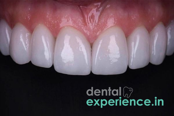
Laminate Veneers: Ultra-thin, custom-designed porcelain shells bonded to the front surfaces of the teeth with minimal surface preparation for maximum stain resistance and natural translucency.
Full-Coverage Ceramic Crowns: Used when older discolored teeth are severely worn, brittle, or structurally compromised to restore long-term aesthetics and function.
Dental treatment costs in Kerala vary depending upon the type of restorative material chosen, facility accreditation, and clinician expertise:
In-Office Professional Bleaching: ₹8,000 – ₹18,000 per session
Direct Composite Veneers: ₹2,500 – ₹5,500 per tooth
Porcelain / E-Max Veneers: ₹9,000 – ₹20,000 per tooth
All-Ceramic / Zirconia Crowns: ₹8,000 – ₹22,000 per crown
Financial Note: Purely cosmetic whitening treatments are generally not reimbursed under standard government health programs or private health insurance plans.
General Dentist: Performs preliminary screenings, professional ultrasonic cleanings, surface stain removal, and standard bleaching procedures.
Prosthodontist: Recommended for complex aesthetic reconstruction, full-mouth rehabilitation, custom ceramic veneers, and placement of crowns on teeth worn down by age or attrition.
Endodontist: Consulted if a single tooth has darkened due to nerve death or pulpal necrosis, requiring internal root canal bleaching, endodontic therapy, or both.
Periodontist: Advisable when age-related tooth darkening is accompanied by significant gum recession, root exposure, or advanced periodontal bone loss.
Immediate / Emergency (Within 2 – 4 Hours): Severe pain, facial swelling, trauma, or a visible tooth fracture/crack.
High Priority / Urgent (Within 24 – 48 Hours): Rapid darkening of a single tooth to black or dark gray following a minor injury.
Elective / Scheduled (At Your Leisure): Generalized, gradual darkening across teeth over many years poses no acute risk and can be addressed based on personal aesthetic preferences.
Why do my teeth appear to darken in color, even though I brush at least two times a day?
Brushing removes surface-level plaque and debris, but the natural aging process causes inner secondary dentin to thicken while the outer enamel thins over time. As the darker dentin layer becomes more visible through the thinned enamel, teeth naturally display a darker shade regardless of brushing frequency.
Can home-based natural products or charcoal powder help restore white color to aged, discolored teeth?
No. Charcoal powders, baking soda, and other coarse abrasives scratch and weaken already thinned tooth enamel. This accelerated enamel loss exposes the underlying yellowish dentin even faster, making teeth look darker over time. Consult a dentist for safe professional whitening or restorative options.
Is professional whitening safe for older teeth with thin enamel?
Yes, provided it is performed or monitored by a qualified dentist. Clinicians evaluate enamel thickness prior to treatment and can apply remineralizing agents along with desensitizing protocols to protect the pulp and minimize post-treatment sensitivity.
Tooth color changes are a normal part of the aging process. Modern restorative treatments aim to optimize aesthetics while preserving as much of the natural tooth structure as possible. Before initiating major cosmetic interventions, patients should undergo a comprehensive clinical evaluation assessing pulp vitality and enamel thickness to select conservative, minimally invasive options. Routine preventive dental visits and consistent oral hygiene practices remain the foundation for maintaining both dental function and smile aesthetics throughout the senior years.



.jpg)
.jpg)
.jpg)
.jpg)
.jpg)
.jpg)
.jpg)
.jpg)
.jpg)
.jpg)
.jpg)
.jpg)