.jpg)
പല്ലുകളിലെ കേടുകൾ അടയ്ക്കാൻ ഉപയോഗിക്കുന്ന കോമ്പോസിറ്റ്, അമാൽഗം, സിമന്റ് തുടങ്ങിയ ഫില്ലിംഗുകൾക്ക് കാലക്രമേണ ഉണ്ടാകുന്ന നിറംമാറ്റത്തെയാണ് 'ഫില്ലിംഗിനുണ്ടാകുന്ന നിറംമാറ്റം' എന്ന് വിളിക്കുന്നത്. ഫില്ലിംഗിന്റെ ഉപരിതലത്തിൽ കറകൾ അടിയുന്നത് കൊണ്ടോ, ഫില്ലിംഗിന്റെയും പല്ലിന്റെയും സന്ധിയിലുണ്ടാകുന്ന സൂക്ഷ്മ വിള്ളലുകളിലൂടെ (Marginal microleakage) ഉമിനീരും ബാക്ടീരിയകളും ഉള്ളിലേക്ക് കടക്കുന്നത് കൊണ്ടോ, ഫില്ലിംഗ് മെറ്റീരിയലുകൾക്ക് ഉണ്ടാകുന്ന രാസമാറ്റം കൊണ്ടോ ഇത് സംഭവിക്കാം. ഈ വിള്ളലുകൾ ഫില്ലിംഗിന് താഴെ വീണ്ടും കേടുകൾ (Secondary caries) രൂപപ്പെടാൻ കാരണമാവുകയും പല്ലിന്റെ ഇനാമലിനെയും ഡെന്റിനെയും നശിപ്പിക്കുകയും ചെയ്യും.
ക്ലിനിക്കൽ നിർവ്വചനം: ബാഹ്യ കറകൾ, വശങ്ങളിലെ ചോർച്ച (Microleakage) അല്ലെങ്കിൽ മെറ്റീരിയലിന്റെ തേയ്മാനം മൂലം ഫില്ലിംഗ് ചെയ്ത ഭാഗങ്ങളിലുണ്ടാകുന്ന നിറംമാറ്റവും ബലക്ഷയവും.
ഉടൻ ഡോക്ടറെ കാണേണ്ട ലക്ഷണങ്ങൾ: ഫില്ലിംഗിന് താഴെ കറുത്ത തണൽ പോലെ കാണുക, ഭക്ഷണം കടിക്കുമ്പോൾ ഉണ്ടാകുന്ന കടുത്ത വേദന, പെട്ടെന്നുണ്ടാകുന്ന സെൻസിറ്റിവിറ്റി, മോശം രുചിയോ വായ്നാറ്റമോ അനുഭവപ്പെടുക.
ചികിത്സാ മാർഗ്ഗങ്ങൾ: ഫില്ലിംഗ് പോളിഷ് ചെയ്യൽ, സീലന്റുകൾ പുരട്ടൽ, ഫില്ലിംഗ് മാറ്റി പുതിയ കോമ്പോസിറ്റ് ചെയ്യൽ, ഇൻഡൈറക്റ്റ് സെറാമിക് വെനീറുകൾ, സിർക്കോണിയ/സെറാമിക് ക്രൗണുകൾ.
കാഴ്ചയിലെ മാറ്റങ്ങൾ: ഫില്ലിംഗ് ചെയ്ത ഭാഗത്തിന്റെ വശങ്ങളിൽ (Margins) സാവധാനം ഉണ്ടാകുന്ന മഞ്ഞ, ബ്രൗൺ അല്ലെങ്കിൽ കറുപ്പ് നിറം.
തൊടുമ്പോൾ അറിയുന്നത്: ഫില്ലിംഗിന് മുകളിലൂടെ നാക്ക് കൊണ്ട് തടവുമ്പോൾ പരുപരുപ്പോ കുഴികളോ അനുഭവപ്പെടുക.
വശങ്ങളിലെ കറകൾ: പല്ലിന്റെ സ്വാഭാവിക ഇനാമലും ഫില്ലിംഗും ചേരുന്ന ഭാഗത്ത് വ്യക്തമായി കാണുന്ന കറുത്ത വരകൾ.
താപനിലയോടുള്ള സെൻസിറ്റിവിറ്റി: ചൂടുള്ളതോ തണുത്തതോ മധുരമുള്ളതോ ആയ ഭക്ഷണങ്ങൾ കഴിക്കുമ്പോൾ ഉണ്ടാകുന്ന ചെറിയ വേദന.
പല്ലിനുള്ളിലെ കറുപ്പ്: പല്ലിന്റെ ഇനാമലിന് താഴെ ചാരനിറത്തിലോ കറുപ്പുനിറത്തിലോ കാണുന്ന തണൽ (ഫില്ലിംഗിന് താഴെ കേട് പടരുന്നതിന്റെ ലക്ഷണം).
ഉപരിതലത്തിലെ പാടുകൾ കേവലം സൗന്ദര്യപ്രശ്നം മാത്രമാണെങ്കിലും താഴെ പറയുന്ന ലക്ഷണങ്ങൾ കണ്ടാൽ അടിയന്തിരമായി ഡോക്ടറെ കാണേണ്ടതാണ്:
വിട്ടുമാറാത്ത മിടിക്കുന്ന വേദന: നാഡിയിലേക്ക് അണുബാധ പടരുന്നതിന്റെ (Irreversible Pulpitis) ലക്ഷണം.
ദുർഗന്ധവും മോശം രുചിയും: ഫില്ലിംഗിന് അടിയിൽ ഭക്ഷണം കുടുങ്ങി ബാക്ടീരിയകൾ വളരുന്നത് മൂലം ഉണ്ടാകുന്ന വായ്നാറ്റം.
പല്ലിന് ഇളക്കമോ പൊട്ടലോ വരിക: ഭക്ഷണം ചവയ്ക്കുമ്പോൾ പല്ല് ഇളകുന്നതായോ ഫില്ലിംഗ് ഭാഗികമായി പൊട്ടിപ്പോകുന്നതായോ തോന്നുക.
മോണയിൽ പഴുപ്പ് കുരു: നിറംമാറിയ ഫില്ലിംഗിന് സമീപത്തെ മോണയിൽ കാണുന്ന ചെറിയ കുരുക്കൾ (Fistula/Parulis) വേരിന്റെ അറ്റത്തെ അണുബാധയെ കാണിക്കുന്നു.
വശങ്ങളിലെ സൂക്ഷ്മ ചോർച്ച (Marginal Microleakage): പല്ലും ഫില്ലിംഗും തമ്മിലുള്ള പറ്റിപ്പിടുത്തം കാലക്രമേണ അയയുകയും ആ ഇടയിലൂടെ ഉമിനീരും ബാക്ടീരിയകളും ഉള്ളിലേക്ക് കടക്കുകയും ചെയ്യുന്നത്.
കറയുള്ള ഭക്ഷണങ്ങൾ: സ്ഥിരമായി കടും നിറമുള്ള ഭക്ഷണങ്ങളും പാനീയങ്ങളും കഴിക്കുന്നത് മൂലം ഫില്ലിംഗിന്റെ പുറംപാളിയിൽ കറകൾ അടിയുന്നത്.
മെറ്റീരിയലിന്റെ രാസമാറ്റം: കോമ്പോസിറ്റ് ഫില്ലിംഗുകൾക്ക് കാലപ്പഴക്കം കൊണ്ട് നിറംമാറുന്നു; അമാൽഗം (വെള്ളി) ഫില്ലിംഗുകൾക്ക് തുരുമ്പെടുക്കൽ (Corrosion) മൂലം ചാര അല്ലെങ്കിൽ കറുപ്പ് നിറം വരുന്നു.
ഫില്ലിംഗിന് താഴെയുള്ള കേടുകൾ (Secondary Caries): പഴയ ഫില്ലിംഗിന്റെ വശങ്ങളിലൂടെ ബാക്ടീരിയകൾ കടന്ന് പല്ലിനുള്ളിൽ വീണ്ടും കേടുകൾ ഉണ്ടാക്കുന്നത്.
പല്ലിനുണ്ടാകുന്ന ആഘാതങ്ങൾ: കടിക്കുമ്പോഴുണ്ടാകുന്ന അമിത മർദ്ദം മൂലം ഫില്ലിംഗിൽ ഉണ്ടാകുന്ന സൂക്ഷ്മ വിള്ളലുകൾ.
കട്ടൻ ചായ, കാപ്പി, മഞ്ഞൾ ചേർത്ത നാടൻ കറികൾ എന്നിവ പതിവായി കഴിക്കുന്നത്.
സിഗരറ്റ്, പുകയില, വെറ്റില-മുറുക്ക് എന്നിവയുടെ ഉപയോഗം.
വായ ശുചിയായി സൂക്ഷിക്കാതിരിക്കുന്നത് മൂലം ഫില്ലിംഗിന് ചുറ്റും പ്ലേക്ക് അടിയുന്നത്.
ഉറക്കത്തിൽ പല്ലുകടിക്കുന്ന ശീലം (Bruxism) മൂലം ഫില്ലിംഗുകൾക്ക് വിള്ളൽ വീഴുന്നത്.
5 മുതൽ 7 വർഷത്തിൽ കൂടുതൽ പഴക്കമുള്ള കോമ്പോസിറ്റ് ഫില്ലിംഗുകൾ.
കണ്ണാടി പരിശോധന: നല്ല വെളിച്ചത്തിൽ കണ്ണാടി നോക്കി ഫില്ലിംഗ് ചെയ്ത ഭാഗത്തിന് ചുറ്റും കറുത്ത വരകളോ നിറവ്യത്യാസമോ ഉണ്ടോ എന്ന് നോക്കുക.
തൊട്ടുനോക്കിയുള്ള പരിശോധന: നാക്കിന്റെ തുമ്പോ വൃത്തിയുള്ള വിരലോ ഉപയോഗിച്ച് ഫില്ലിംഗിന് മുകളിലൂടെ തടവി നോക്കുക. പല്ലും ഫില്ലിംഗും ചേരുന്ന ഭാഗം മിനുസമുള്ളതായിരിക്കണം; കൂർത്ത അരികുകളോ കുഴികളോ ഉണ്ടെങ്കിൽ ഫില്ലിംഗിന് തകരാറുണ്ട്.
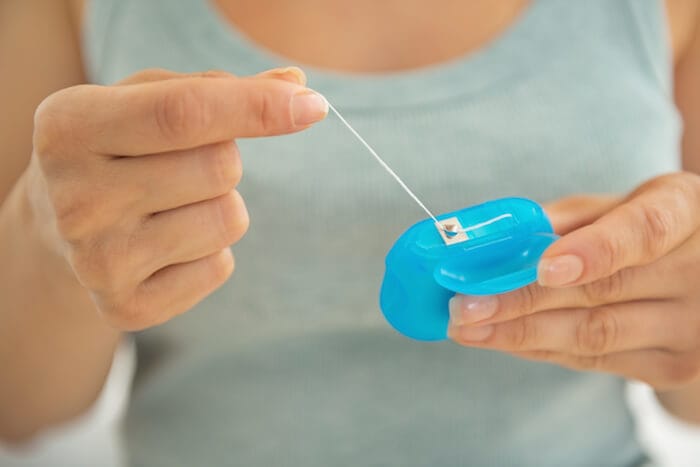
ഭക്ഷണം കുടുങ്ങുന്നത് ശ്രദ്ധിക്കുക: ഫില്ലിംഗ് ചെയ്ത ഭാഗത്ത് വിട്ടുമാറാതെ ഭക്ഷണം കുടുങ്ങുകയോ ഫ്ലോസ് ഉപയോഗിക്കുമ്പോൾ നൂൽ കീറിപ്പോവുകയോ ചെയ്യുന്നുണ്ടോ എന്ന് നോക്കുക.
തണുത്ത വെള്ളം ഉപയോഗിച്ചുള്ള ടെസ്റ്റ്: തണുത്ത വെള്ളം കുടിക്കുമ്പോൾ പെട്ടെന്ന് മാറിപ്പോകുന്ന സെൻസിറ്റിവിറ്റിയാണോ അതോ നീണ്ടുനിൽക്കുന്ന കടുത്ത വേദനയാണോ എന്ന് ശ്രദ്ധിക്കുക.
പ്രധാന മുന്നറിയിപ്പ്: പരിശോധിക്കാൻ കൂർത്ത ലോഹ വസ്തുക്കളോ ടൂത്ത്പിക്കോ പിന്നുകളോ ഉപയോഗിച്ച് ഫില്ലിംഗിന്റെ അരികുകളിൽ കുത്തരുത്; ഇത് ഇനാമൽ പൊട്ടിപ്പോകാനും ഫില്ലിംഗ് ഇളകിപ്പോകാനും ഇടയാക്കും.
To evaluate a discolored dental restoration, a dentist will perform a series of clinical diagnostic assessments:
Tactile and Visual Examination: Using a dental mirror and a blunt CPI probe or light tactile assessment to check restoration margins for catches, micro-gaps, or softened tooth structure indicative of secondary decay without damaging the adhesive seal.
Digital Radiography: Intraoral bitewing and periapical radiographs help evaluate restoration contours, margin fit, and secondary caries beneath existing fillings.
Fiber-Optic Transillumination (FOTI): Directing a specialized fiber-optic light through the tooth allows the clinician to visualize internal micro-fractures, structural cracks, and demineralized carious shadows beneath the restoration.
Pulp Vitality Testing: Thermal testing (cold/heat) or electric pulp testing (EPT) is conducted to determine whether the pulp tissue is vital, inflamed, or necrotic.
|
Severity Level |
Clinical Characteristics |
Recommended Intervention |
|
Grade 1: Superficial Staining |
External surface staining without marginal gaps, microleakage, or discomfort. |
Professional polishing & surface refinishing. |
|
Grade 2: Marginal Degradation |
Visible dark border at restoration edges; mild temperature sensitivity; surface roughness. |
Marginal seal repair or direct composite replacement. |
|
Grade 3: Deep Microleakage & Secondary Decay |
Extensive discoloration, secondary carious lesion beneath filling, moderate localized pain. |
Complete excavation of decay & placement of a composite restoration or indirect ceramic inlay/onlay. |
|
Grade 4: Pulpal Involvement |
Severe darkening, extensive structural loss, spontaneous/continuous throbbing pain, or periapical abscess formation. |
Endodontic root canal therapy followed by post-and-core and full-coverage crown restoration. |
Polishing and Refining (Extrinsic Stains): Polishing the surface of the composite filling using fine diamond burs and specialized polishing pastes to remove extrinsic surface staining.
Composite Filling Replacement: The old filling material is completely removed, the cavity preparation is disinfected, and a new nano-hybrid composite resin is bonded in place, shade-matched to your natural teeth.
Ceramic Indirect Inlays/Onlays: Custom porcelain or ceramic restorations fabricated by dental laboratories that provide superior stain resistance and structural support for larger areas needing restoration.
Full Ceramic Crowns: For teeth with extensive structural wear, severe decay, or dark internal discoloration, a full-coverage zirconia or e.max ceramic crown protects and restores the form, function, and aesthetics of the tooth.
The cost of services varies depending upon the provider's credentials, the materials used, and their location in Kerala:
Polish / Refinish Services: ₹800 – ₹1,500
Nano-Hybrid Composite Replacement (per tooth): ₹1,500 – ₹3,500
Ceramic Inlay/Onlay (per tooth): ₹6,000 – ₹12,000
e.max / Zirconia Ceramic Crown (per tooth): ₹8,000 – ₹18,000
Insurance coverage varies from one plan to another: Cosmetic polishing and refinishing are excluded from most private health and dental insurance plans in India. However, if a patient requires restorative treatment due to trauma or symptomatic secondary caries, partial reimbursement may be available under dental insurance riders. Most state government employee health insurance schemes cover basic clinical restorations and extractions at empanelled hospitals, while aesthetic or ceramic upgrade costs are typically paid out-of-pocket by patients.
General Dentist (GDP): Qualified to identify failing restorations, routinely replace fillings with composite resin, and perform routine dental prophylaxis (cleanings).
Prosthodontist: Recommended when you require complex aesthetic replacements, full-mouth reconstruction, or custom ceramic crowns and veneers.
Endodontist: Needed when deep secondary decay reaches the dental pulp, requiring root canal treatment in addition to the removal of the compromised filling.
Immediate (Within 24 Hours): Severe sharp pain radiating through the face, visible facial swelling, or a dislodged filling that leaves an exposed cavity.
Prompt Visit (Within 3–5 Days): Sharp pain when chewing or biting down, a loose or disintegrating filling, or persistent food impaction between teeth.
Elective Visit (Within 2–4 Weeks): Mild superficial staining or progressive aesthetic discoloration without pain, or routine cosmetic smile upgrades.
What causes a composite tooth filling to darken in color over a period of a couple of years?
Dark food and beverage colors—such as coffee, tea, and spices like turmeric—will slowly stain a composite filling because resin matrices naturally absorb surface pigments over time. Furthermore, if marginal microleakage develops around an older composite filling, it allows oral bacteria, fluids, and debris to enter the gap, causing internal discoloration and secondary staining.
Can I use bleaching gel to whiten a stained composite tooth filling?
No. Whitening agents and bleaching gels only work on natural tooth enamel. Restorative materials (such as composite resin, porcelain, and amalgam) do not respond to whitening agents. To restore their original brightness, the filling must either be professionally polished by your dentist or replaced entirely.
How can I keep my new composite filling looking clean and white?
Keep your teeth and gums clean by brushing at least twice a day using a soft-bristled toothbrush and flossing daily. Limit your intake of dark-colored beverages and staining foods. Rinse your mouth out with water immediately after consuming foods or drinks containing turmeric. Avoid smoking and tobacco products, and visit your dentist every six months for routine professional cleanings and check-ups.
The discoloration of a restoration is more than just a cosmetic issue. A stained margin from a composite resin restoration is usually an indication that marginal microleakage is occurring at that site, which in turn can lead to secondary caries. Therefore, early evaluation with digital radiography allows for the conservative replacement of a failing restoration, preserving the original tooth structure and preventing invasive endodontic treatment or complex restorative coverage.






.jpg)
.jpg)
.jpg)
.jpg)
.jpg)
.jpg)
.jpg)
.jpg)
.jpg)
.jpg)
.jpg)
.jpg)